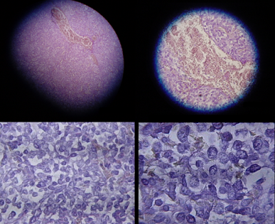
Immunohistochemical studies play an important role for the pathologist in differentiating malignant mesothelioma from neoplastic mimics, such as breast or lung cancer that has metastasized to the pleura.
 |
| Micrographs showing mesothelioma in a core biopsy. |
There are numerous tests and panels available, but no single test is perfect for distinguishing mesothelioma from carcinoma or even benign versus malignant. The positive markers indicate that mesothelioma is present; if other markers are positive it may indicate another type of cancer, such as breast or lung adenocarcinoma.
Calretinin is a particularly important marker in distinguishing mesothelioma from metastatic breast or lung cancer.
Typical immunohistochemistry results
 |
| Typical immunohistochemistry results |
Subtypes
There are three main histological subtypes of malignant mesothelioma: epithelioid, sarcomatous, and biphasic. Epithelioid and biphasic mesothelioma make up approximately 75-95% of mesotheliomas and have been well characterized histologically, whereas sarcomatous mesothelioma has not been studied extensively. Most mesotheliomas express high levels of cytokeratin 5 regardless of subtype.
Epithelioid mesothelioma is characterized by high levels of calretinin.
Sarcomatous mesothelioma does not express high levels of calretinin.
 |
| Micrograph of a pleural fluid cytopathology specimen showing mesothelioma. |
Other morphological subtypes have been described :
- Desmoplastic
- Clear cell
- Deciduoid
- Adenomatoid
- Glandular
- Mucohyaline
- Cartilaginous and osseous metaplasia
- Lymphohistiocytic
Differential diagnosis :
- Metastatic adenocarcinoma
- Pleural sarcoma
- Synovial sarcoma
- Thymoma
- Metastatic clear cell renal cell carcinoma
- Metastatic osteosarcoma

No comments:
Post a Comment